AI-Based biological cells (RGC) / Materials grain size analysis
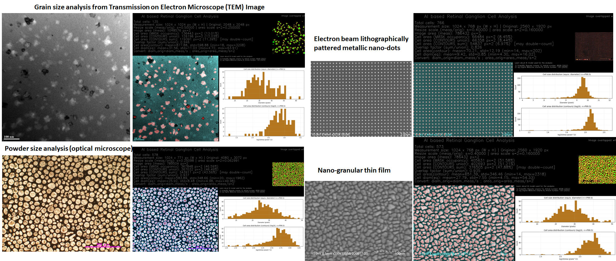
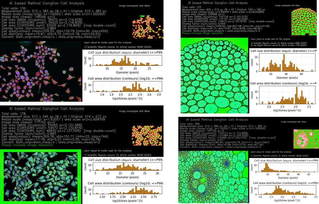
Upload a fluorescence microscopy image (e.g., RGC / nuclei / cells) or a granular / microstructure image (SEM, TEM, metallography, powders, nanocrystalline materials, stones, rice, wheat, pulses, etc.). (Last updated: 1 March 2026)
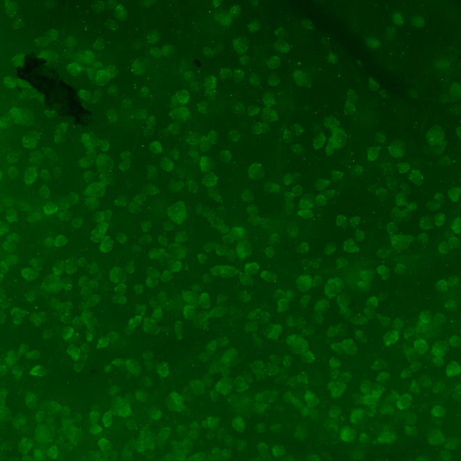
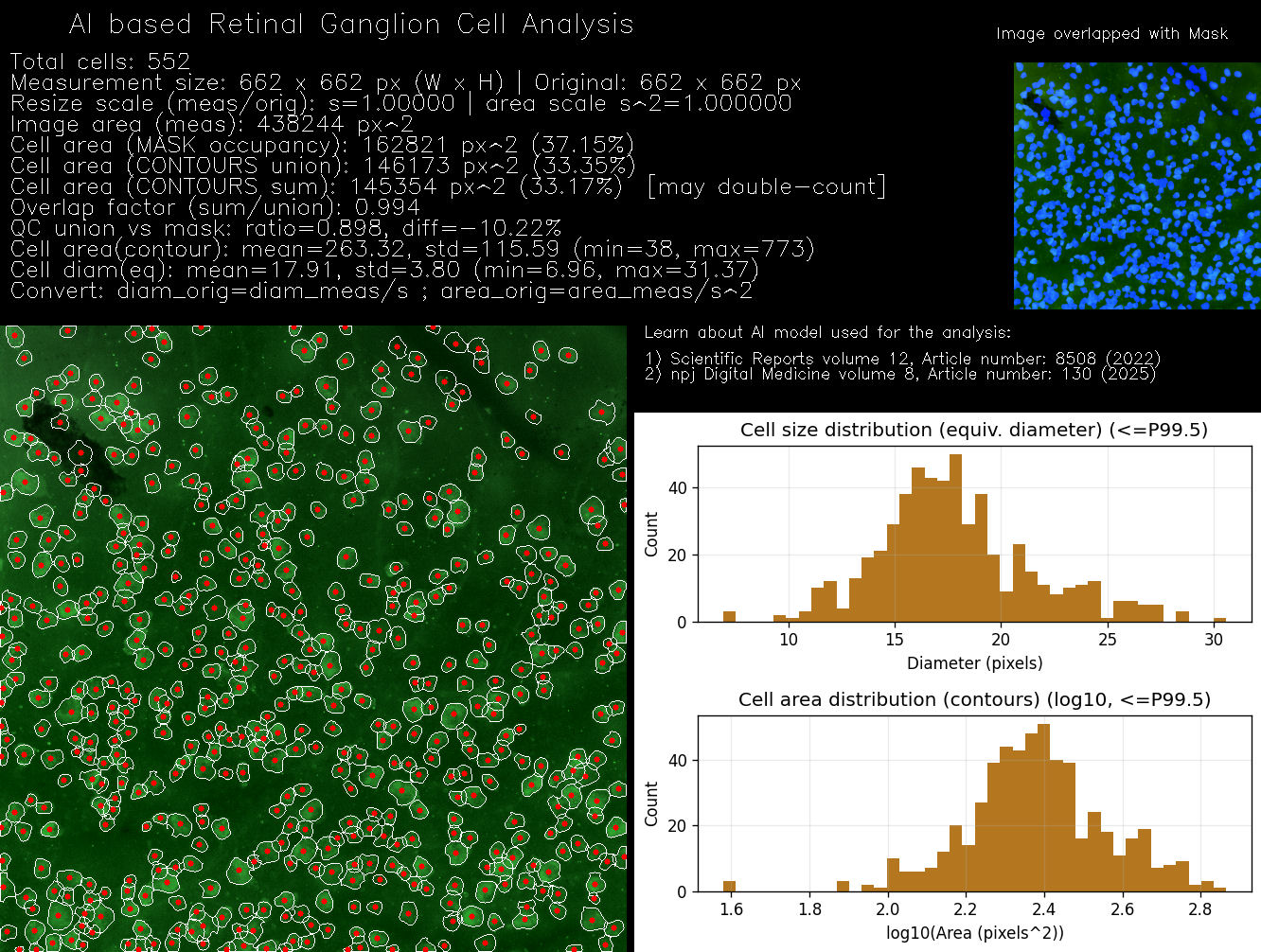
Retinal ganglion cells (RGCs) exist in all vertebrate retinas, including humans and mice. They are the output neurons that transmit visual information to the brain via the optic nerve. Quantitative analysis of RGC loss is a standard endpoint in glaucoma and optic nerve injury research.
This research demonstration supports AI-based detection, segmentation, and counting for biological cells (e.g., RGC fluorescence microscopy) and grain/particle segmentation and size statistics for materials images (e.g., SEM/TEM). Please upload only images that you are permitted to use.